Previous
Next 
Separation principles
Description
This SCO will show you the principles of separation used.

Principles

The most known separation process is filtering. In any common filtering process, the analyte mixture is migrated through a substance to filter the mixture. For most proteomics and metabolomics applications however, filtering is too restrictive, as there is only passed or not passed molecules.
The key principle for a better resolution of separation is retention in contrast to blocking as in a filter. If the speed of migration of the analyte through some substance was influenced by some physical property differently strong in different molecule entities in the analyte, one gets (continuous) separation over time via the distance of migration. 
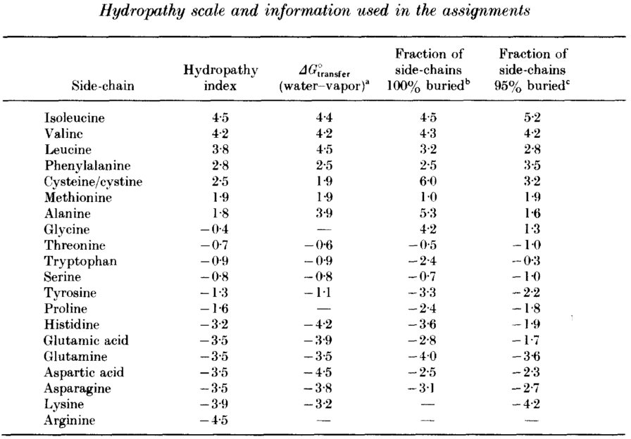
Image by Magnus Manske ( Creative Commons Attribution-Share Alike 3.0 )
The principle of migrating an analyte through a substance is applied in many separation techniques, e.g. in PolyAcrylamide Gel Electrophoresis (PAGE). An electric current is applied to drive the analyte from a loading pocket through the gel.  
Teh application of an electric current for separation is called electrophoresis. The migration speed is inversely proportional to the net charge and the hydrodynamic volume. Depending on the mode of PAGE though, separation can be function of different properties.
  • Without any detergent, the separation is a function of the net charge of the native molecule and the size and shape of the molecule. This is known as Native-PAGE
  • In SDS-PAGE, the analyte may be treated with an detergent agent like sodium dodecyl sulfate (SDS), not only bringing proteins into linear form, but also applying a uniformly placed number of charges over the length of the protein. This will also render the analyte molcules almost unusable for downstream technologies. The separation is a function dominated by the charge. And hence the charges are approximately uniformly distributed over the molecules size, on a rough scale, it can also be seen as a function of molecular mass.
There are, apart from the obvious size, shape, and mass (making up the hydrodynamic volume), two more physical properties of the analytes, that provide the grounds for good separation. They get employed in important separation techniques that will be introduced in the next chapter. The most important will be introduced in the following.

The isoelectric point

The isoelectric point (\(pI\)) of proteins is the \(pH\) value of a solution at which the molecules positive and negative charges are in balance.
Following the definition of Brønsted-Lowry, proton donors are called acids and protein acceptors are called bases.
Proteins, though built from amino acids, have both basic and acidic properties (also called Zwitterionen). This comes from the various side chains of amino acids having various \(pK_a\) values. The acid dissociation constant \(K_a\) is commonly given in a negative logarithmic scale \(pK_a\) due to the many orders of magnitude spanned. The larger \(K_a\) is, the more dissociation is taking place, thus the stronger the acid in solution (more protons donated following the Brønsted-Lowry scheme).
Like this, proteins can be separated according to their isoelectric point using a technique called isoelectric focusing. It uses a \(pH\) gradient to separate proteins on a gel, and also the first step in 2-D gel separation.
The \(pI\) of a protein can be calculated or experimentally determined with isoelectric focusing.
The theoretical calculation can be done by considering all free ionizable groups. These are the N-terminal amino group, the C-terminal carboxyl group and the ionizable sidechains:
  • Asp (D)
  • Glu (E)
  • Arg (R)
  • Lys (K)
  • His (H)
  • Cys (C) (-SH)
  • Tyr (Y)
Histidine (H), lysine (K), and arginine (R) carry a proton acceptor group in their side chains.
Cysteine (C), aspartate (D), glutamate (E), and tyrosine (Y) have proton donor groups in their side chains.
Note that the \(pK\) values for the termini also vary with the actual amino acids that form the corresponding ends.
Using the dissociation constants for these ionizable groups we can calculate a estimation of the pI of a protein. The partial charges are calculated for each amino acid x at any given \(pH\) is:
\(C_x = 1/(1 + 10^{pH-pK_x})\)
if the amino acid is positively charged, and
\(C_x = -1/(1 + 10^{pK_x-pH})\)
if the amino acid is negatively charged.
A function \(C_P(pH)\) can now estimates the net charge of a protein \(P\) for a given \(pH\) with:
\(C_P(pH) = \sum_{x in P}{C_x}\)
The protein's \(pI\) is \(C_P(pH) = 0\).

Here is an algorithm that calculates the pI of a given sequence of amino acids:
const: \(\epsilon\) limit for accuracy
var: \(x,x_0,x_1\)
begin
  • find values \(x_0, x_1\) that \(sign(C(x_0)) = sign(C(x_1))\)
  • while [\(x_1 - x_0\)] \(\geq \epsilon\) and max number of iterations not reached do
    • \(x := (x_0 + x_1)/2\)
    • if \(sign(C(x_0)) \neq sign(C(x))\) then \(x_1 := x\) else \(x_0 := x\) end
  • end
end
\(pI\) is in [\(min(x_0, x_1), max(x_0, x_1)\)]
EXERCISE: calculate the isolelectric point of the peptide SYFPEITHI.
Hydrophobicity

Compounds that repel water or any other polar solvent are called hydrophobic. They do not easily dissolve in water, rather tend to stick together. This is called a hydrophobic interaction. This interaction is entropy driven, as forming a 'cage' of polar molecules around the hydrophobic molecules is reducing entropy. It is reducing the interface between both. But if interactions between hydrophobes will occur it will increase entropy, disturbing the 'cage' of molecules around the hydrophobes, increasing enthalpy \(\Delta H\) (breaking H-Bonds in the polar solvent to some small extent), and increase entropy \(\Delta S\) (to a greater extent).
In terms of the Gibbs free energy:
\(\Delta G = \Delta H - T \Delta S\)
This implies, that if \(\Delta G\) is negative, hydrophobic interactions are favoured and will happen spontaneous, if \(\Delta S\) and \(\Delta H\) are positive. (Assumed temperature \(T\) is constant).
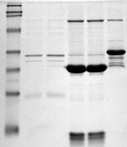
The hydrophobicity of amino acids is determined via experiments or calculations. There are several different measures, the most common is the Kyte-Doolittle scale, where most hydrophobic residues are given highest positive scores (Kyte, J. and Doolittle, R. 1982. A simple method for displaying the hydropathic character of a protein. J. Mol. Biol. 157: 105-132).
A hydrophobicity value for a protein can be calculated simply by adding the hydrophobicity values of all the residues and dividing the sum by the number of residues. This is called a GRAVY score (GRand AVerages of hYdrophobicity).

EXERCISE: calculate the GRAVY score of SYFPEITHI.
 Previous
Next